flow cytometry results leukemia
Cancer is the result of genetic mutations in specific cells. Flow Cytometry is used in several fields including molecular biology pathology immunology virology plant biology and marine biology.

International Clinical Cytometry Society
HEMATOPATHOLOGY New Diagnosis Follow-up.

. Chronic myelogenous l See. This will depend on the specific types of cells under investigation as well as what lab is used. Beckman Coulter is revolutionizing the leukemia and lymphoma analysis in clinical flow cytometry laboratories with the innovative ClearLLab Solutions.
D Percentages of caspase apoptotic cells in mouse LSCs n. Leukemic cells can also be identified by flow cytometry and immunocytochemistry which rely on antibodies binding to and helping to identify malignant cells. Staining of A549 cells with 5 μM Vybrant DyeCycle Violet results in a poor DNA content histogram because the dye is actively pumped out of.
Flow cytometry can detect certain antigens CD41 CD61 on the blast cells which are typical for AML-M7. CML results from a translocation of genetic. Flow cytometry test results can take up to several weeks to come back.
Here an immune risk score was explored to predict the survival of patients with AML. Some of the common application include. BD Biosciences flow cytometry reagents truly reflect our scientific leadership in flow cytometry innovation and our 45 years of dedication to providing high-quality products.
Calgary Alberta T2N 2T9 -944 4765 Fax. Developed for flow cytometry StarBright Dyes are suitable for resolving dim and rare populations yet flexible enough to fit into any flow cytometry panel. Our comprehensive reagent portfolio includes clinical diagnostic testing kits kits for innovative new approaches to clinical research and single-color reagents for.
Six flow cytometry-validated immune. A human T cell leukemia cell line were pulsed with 10 µM EdU for 2 hours prior to detection with Alexa Fluor 647 azide. Spectra viewers will help you determine the amount of spillover and excitation by each laser.
It is used in clinical labs for the detection of malignancy in bodily fluids like leukemia. An abnormal cell will show different patterns that may suggest the presence of leukemia lymphoma or other diseases. To help you get consistently reproducible results refer to.
C Representative flow cytometry plots and statistics of the percentages of apoptotic cells in mouse LSCs n 3 individual mice. CD4 CD4 Count CD3 CD4 CD8 Lymphocytosis 30x10. They provide the theory and key practical aspects of flow cytometry enabling immunologists to avoid the common errors that often.
Mangaonkar AA et al. 40 and 50 affecting slightly more men than women 4600 adults in the US. Flow cytometers contain three main systemsfluidics optics and electronics.
403 270 4135 CLSFlowCytometryclsabca Shaded areas are Required Information. With AML flow cytometry can help to detect special subgroups. Results from flow cytometry have a quick turnaround often less than one day.
A hematopathologist is a. Flow cytometry DNA content distribution in a cell cycle analysis assay. No current model includes covariates related to immune cells in the AML microenvironment.
Since T-PLL is rare it is important that an experienced hematopathologist examine and interpret the patients lab results. BTW this sounds easier than it is. Your healthcare provider will discuss your flow cytometry results in detail and talk about possible treatment options.
Blood chemistry results can indicate the severity of leukemia as well as how well organs such as the kidneys or liver are functioning. It is for example rather difficult to prove an AML-M7 megakaryoblastic leukemia without flow cytometry. Practical limitations of monocyte subset repartitioning by multiparametric flow cytometry in chronic myelomonocytic leukemia.
IMMUNE MONITORING CLINICALLABORATORY FINDINGS. Flow Cytometry - Foothills Medical Centre 1403-29th Street NW. T-cell prolymphocytic leukemia T-PLL is an extremely rare and typically aggressive malignancy cancer that is characterized by the out of control growth of mature T-cells T-lymphocytes.
These guidelines are a consensus work of a considerable number of members of the immunology and flow cytometry community. Diagnostic Assay Development Clinical Diagnostic Antigens and Antibodies Immunoglobulins Leukemia Markers Tumor Markers. ClearLLab 10C system is an integrated FDA cleared and CE-marked IVD leukemia and lymphomaLL immunophenotyping solution for lymphoid and myeloid lineages using the dry DURA Innovations technology.
These tests are not used to diagnose leukemia. There are useful tools that can help with panel design. Prediction models for acute myeloid leukemia AML are useful but have considerable inaccuracy and imprecision.
The fluidics system funnels a sample of cells eg a sample of human blood into a single stream so that the cells pass through a laser beam one at a time. For more in-depth information on Antibody Titration see our Antibody Titration in Flow Cytometry page. The bone marrow aspirate sample is treated with special antibodies that stick only to the.
How flow cytometry works. As each cell passes through the beam it scatters light and may emit fluorescent light. A fresh bone marrow aspirate sample is required for reliable results.
Flow cytometry is a technique that evaluates individual cells by checking for the presence or the absence of certain protein markers on the cell surface. Abnormal results are usually found in the presence of.
Flow Cytometry Panel Of Cd14 And Cd16 Expression On Gated Monocytes In Download Scientific Diagram

Applications Of Flow Cytometric Immunophenotyping In The Diagnosis And Posttreatment Monitoring Of B And T Lymphoblastic Leukemia Lymphoma Digiuseppe 2019 Cytometry Part B Clinical Cytometry Wiley Online Library

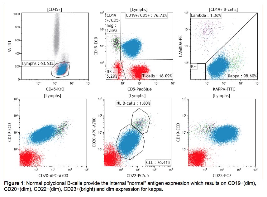
Flow Cytometry And Cell Sorting Strategy For B Cll Cells Cells Are Download Scientific Diagram
Gating Strategy For Cell Counting By Flow Cytometry A Initial Download Scientific Diagram

Flow Cytometric Presentation Of A Large B Cell Lymphoma A Forward Download Scientific Diagram

Flow Cytometric Monitoring For Residual Disease In B Lymphoblastic Leukemia Post T Cell Engaging Targeted Therapies Cherian 2018 Current Protocols In Cytometry Wiley Online Library

Impact Of Flt3 Receptor Cd135 Detection By Flow Cytometry On Clinical Outcome Of Adult Acute Myeloid Leukemia Patients Clinical Lymphoma Myeloma And Leukemia

Verification Of Cdc Or Pdc Depletion By Flow Cytometry Lung Download Scientific Diagram

Flow Cytometry Based Protocols For Human Blood Marrow Immunophenotyping With Minimal Sample Perturbation Star Protocols

A Flow Cytometry Analysis Of Cd44 Cd24 Cell Surface Markers Download Scientific Diagram

5 Easy Steps For Successful Flow Cytometry Bio Rad Flow Cytometry Flow Success

Chapter 7 Some Clinical Applications Flow Cytometry A Basic Introduction

Warde Medical Laboratory Medical School Stuff Medical Laboratory Medical Training

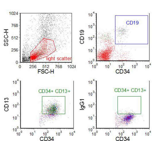
Flow Cytometry The Cd19 Positive Cells Are Positive For Cd5 Cd20 Download Scientific Diagram

Gating Strategies For Effective Flow Cytometry Data Analysis Bio Rad Flow Cytometry Flow Data Analysis

Flow Cytometric Analysis Of Peripheral Blood From A Horse With Acute Download Scientific Diagram

Reproducible Diagnosis Of Chronic Lymphocytic Leukemia By Flow Cytometry An European Research Initiative On Cll Eric European Society For Clinical Cell Analysis Escca Harmonisation Project Rawstron 2018 Cytometry

Immunophenotyping Hscs From Mouse Bone Marrow Protocol Deutschland

Flow Cytometry Analysis Of Ep On Apoptosis And Cell Cycle Progression Download Scientific Diagram